Nonlinear microscopy
How to build a two-photon microscope
Excitation confined to the focus, so no pinhole is needed and scattered light still counts as signal. Everything then follows from one scaling law.
Signal ∝ P²/(τ·f)
Absorption is quadratic in intensity. Shorter pulses and lower repetition rates buy signal at the same average power — and the same heating.
No pinhole
Only the focal volume is excited, so every collected photon is in-focus by definition. Collect non-descanned and keep the scattered light.
Dispersion is the tax
The objective and scan optics stretch the pulse. A 100 fs pulse arriving as 300 fs costs two thirds of your signal.
Interactive excitation-budget lab
Where your two-photon signal actually goes
Pulse at the sample
149 fs
Peak power
1.6 kW
Pulse energy
0.25 nJ
Relative signal
0.67×
Dispersion penalty
100 fs at the laser → 149 fs at the focus · 67% of the signal retained
Duty cycle, focal confinement and dose are in a workable range for this configuration.
Why there is no pinhole
Excitation per z-plane, normalised to the focal plane · NA 1.00 · 920 nm · zR ≈ 389 nm
Widefield
- Excited
- Entire double cone
- Detected
- Everything, in-focus and out
Full-depth bleaching
Confocal
- Excited
- Entire double cone
- Detected
- Pinhole rejects out-of-focus
Full-depth bleaching — the pinhole discards the signal, it does not prevent the damage
Two-photon
- Excited
- Focal volume only
- Detected
- All of it, non-descanned
Confined to the focal plane
The one-photon line is flat because total power is conserved as the beam spreads: every plane above and below the focus absorbs the same amount. That is the photobleaching a confocal pinhole cannot prevent — it only discards the emission afterwards.
A first-order model for comparing configurations, not a substitute for measuring pulse width at the focus. Real systems add higher-order phase, objective transmission that falls steeply past 1000 nm, and wavelength-dependent scattering in tissue.
Femtosecond sourcesThe beam path
Six stages between the laser and the detector
Source
Femtosecond pulses, 80 MHz. Rep rate sets pulse energy at a given average power.
Femtosecond lasersPower control
An AOM sets power per plane and blanks the beam during flyback. Add an isolator to protect the laser.
AOM & shuttersBeam expansion
Fill the objective back aperture. Underfilling lowers the effective NA and throws away resolution and signal.
Beam expandersScan & relay
Scan lens and tube lens conjugate the mirror to the pupil, so the beam pivots without walking off it.
Cage & relay opticsZ and sample
Piezo objective or sample positioning for stacks. Closed-loop if plane spacing must be metrology.
Piezo stagesCollection
Dichroic and short-pass blocking, collected non-descanned. Scattered fluorescence still carries signal.
Filters & dichroicsDetectors for two-photon are PMTs or hybrid detectors placed close to the objective. We do not supply those — for descanned or widefield channels alongside the nonlinear path, see the scientific cameras.
Sources
80 MHz femtosecond lasers from the catalog
Wavelength follows the fluorophore; average power has to survive the scan path before it reaches the sample.
FS-H-780A
780 nm
GFP, CFP and DAPI-class fluorophores sit near their two-photon peak around 780 nm.
- Output / average power
- 1-50 mW
- Repetition rate
- 80±2 MHz
- Single pulse energy (nJ)
- 0.1-0.6
FS-H- 1030B
1030 nm
Longer wavelengths scatter less. Headroom for power lost to the scan path and the objective.
- Output / average power
- 500-3000 mW
- Repetition rate
- 80±2 MHz
- Single pulse energy (nJ)
- 6.25-30
FS-H- 10 64B
1064 nm
The most average power of the three, for thick specimens and multi-point excitation.
- Output / average power
- 500-5000 mW
- Repetition rate
- 80±2 MHz
- Single pulse energy (nJ)
- 6.25-62.5
Before you order
Six things that decide whether it works
- Measure pulse width at the focus, not at the laser head — the objective is usually the largest dispersive element.
- Check objective transmission at your wavelength; many drop steeply beyond 1000 nm even when nominally IR-rated.
- Confirm the scan lens and tube lens conjugate the scan mirror to the back pupil, or the beam walks off at large field angles.
- Fill, but do not heavily overfill, the back aperture — overfilling wastes power for marginal resolution.
- Budget average power for heating and pulse energy for photodamage separately. They fail in different ways.
- Class 4 invisible beam: enclose the path and specify goggles rated at the excitation wavelength.
Before you specify
Guides that cover this decision
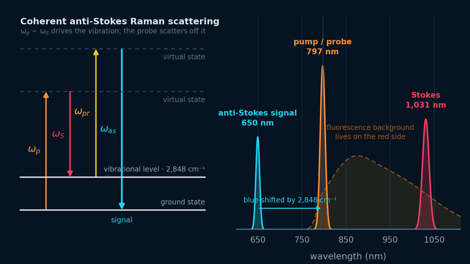
Coherent Raman imagingBuilding a CARS microscope
Coherent anti-Stokes Raman scattering, from the four-wave-mixing physics to the hardware: excitation schemes, pulse width versus linewidth, the delay line that carries the spectral axis, and the detection chain.
Open guide Microscopy
How to choose a laser for microscopy
How much power each imaging modality actually needs at the sample, which excitation lines match your fluorophore panel, why modulation bandwidth beats raw milliwatts, and the photon count above which laser noise finally reaches the image.
Open guide Ultrafast spectroscopy
Building a pump–probe spectroscopy system
Design the pump, probe, delay line, monochromator, visible or SWIR camera, synchronization, and transient-signal budget as one experiment.
Open guide Confocal microscopy
Designing a confocal fluorescence excitation system
Connect fluorophores, pinhole size, Airy units, spatial sampling, scan timing, modulation, and power after the fiber in one practical design workflow.
Open guideSpecify the system
Tell us the fluorophore and the depth.
We will size the wavelength, power and dispersion budget with you before anything is quoted.