
Light-sheet microscopy
SPIM, DSLM and mesoSPIM
Illuminate one plane, image it from the side. Every design decision after that comes from a single geometric constraint.
w₀ = λ / (π·NA)
Illumination NA sets the waist. That is the only knob that makes a sheet thin.
z_R ∝ 1 / NA²
It also sets how far the sheet stays thin. Halve the thickness, quarter the field.
Dose ∝ planes
Only the imaged plane is lit, so a stack costs one exposure per plane instead of one per plane per plane.
Interactive sheet-geometry lab
A thinner sheet is always a shorter sheet
Start from an instrument
Sheet thickness
7.32 µm
Sheet stays thin across
661 µm
Camera field
666 µm
Detection DOF
1.01 µm
Sheet profile across the frame
Sheet covers 99% of the imaged field
The sheet covers the frame, and at 7.3 µm against a 1.01 µm depth of field it is the sheet — not the objective — that sets your optical section. Standard for a full-field light sheet; raise illumination NA only if you are willing to tile.
Gaussian-sheet approximation in water (n = 1.33). Bessel and Airy sheets trade side lobes for a longer field; axially swept (ASLM) and tiled approaches break the tradeoff at the cost of speed or complexity.
sCMOS detectionThree points on one curve
The same optics at different scales
SPIM
Cells to embryos · µm sheet
A cylindrical lens forms a static sheet. Simplest to build; intensity varies across the sheet and stripes appear behind absorbing structures.
DSLM
Same scale, cleaner field
A galvo sweeps a pencil beam to synthesise the sheet. Uniform illumination, dual-side and pivot options, and the scan can be synced to a rolling shutter.
mesoSPIM
Cleared organs · mm field
Very low illumination NA over a centimetre-scale specimen. The sheet is tens of µm thick, so sectioning comes from the geometry, not from a thin waist.
The chain
Illumination arm, sample, detection arm
Multi-line source
One fiber-coupled head carrying every excitation line, with per-line power control.
Multi-wavelength lasersCollimation
A fiber collimator sets the beam diameter that the illumination optics turn into sheet NA.
Fiber collimatorsSheet forming
A cylindrical lens for static SPIM, or a galvo and scan lens for DSLM. This choice sets illumination NA.
Precision opticsSample translation
The specimen moves through the sheet. Plane spacing is only as good as the stage that sets it.
Motorised stagesOrthogonal detection
Detection objective at 90°, with the sheet parked in its focal plane across the whole frame.
Cage & mountsFiltering & camera
Emission filter, then a large-format sCMOS. Sensor size and magnification fix the field you must keep the sheet thin across.
FiltersThe detail that catches people out
Rolling shutter is a feature here, not a defect
An sCMOS exposes row bands in sequence. In most microscopy that is a nuisance. In a scanned light sheet it is free contrast: sweep the beam so the illuminated line tracks the active rows, and every row is read while only its own plane is lit. Scattered light arriving from elsewhere lands on rows that are not listening.
It costs nothing in hardware and it needs the scan waveform locked to the sensor line time. Verify that the camera exposes the line period and accepts an external trigger before you design around it.
Illumination
Multi-line sources from the catalog
RGB-405/488/561/640
The standard 405/488/561/640 set covering DAPI through far-red, at 30 mW per line.
- Operating mode
- CW
- Output / average power
- 30/30/30/30 mW
FC-405/450/488/520/561/637-CH
Fiber delivery decouples the source from the illumination arm — the mode is clean and the head can sit off the table.
- Operating mode
- CW
- Output / average power
- 20/20/20/20/20/20 mW
Detection
Sensor format decides the field
sMAX04BM
2048² back-illuminated sCMOS at 72.5 fps — fast enough that stage motion, not readout, limits your stack rate.
- Sensor
- GSENSE2020BSI (sCMOS)
- Resolution
- 4.2 MP (2048×2048)
- Pixel Size
- 6.5 µm × 6.5 µm
- Frame Rate
- 72.5 fps @ 2048×2048; 72.5 fps @ 1024×1024
sMAX16BM-U3-CL
4096² across a 36.8 mm sensor. At low magnification this is the format that turns a mm-scale sheet into pixels.
- Sensor
- GSENSE4040BSI (sCMOS)
- Resolution
- 16.8 MP (4096×4096)
- Pixel Size
- 9 µm × 9 µm
- Frame Rate
- 20 fps @ 4096×4096 (USB3); 23 fps @ 4096×4096 (Camera Link)
Before you order
Six checks
- Match refractive index end to end — clearing agent, chamber window, and objective immersion. Mismatch adds spherical aberration that no sheet geometry recovers.
- Confirm the detection objective’s working distance clears the chamber and the illumination arm.
- Decide early between one-sided and dual-sided illumination; retrofitting the second arm is rarely cheap.
- Budget stage step accuracy against your plane spacing — the z-axis is your axial sampling.
- Check the camera exposes line period and external trigger if you intend to sync a scanned sheet.
- Size the data rate: 4096² at 20 fps is roughly 670 MB/s sustained. Plan storage before the first stack.
Before you specify
Guides that cover this decision
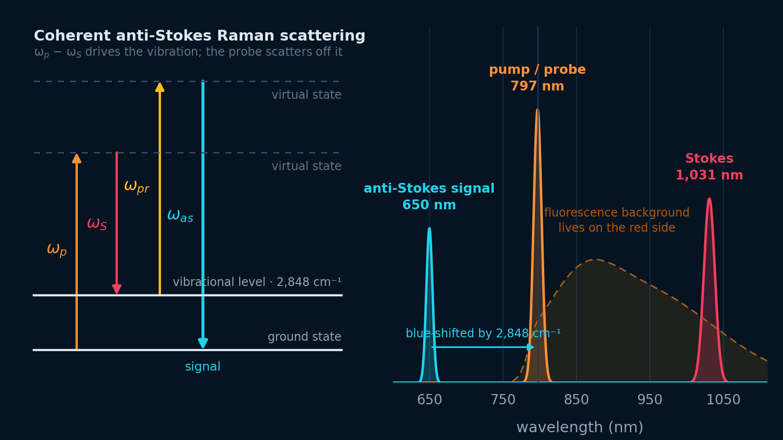 Nonlinear microscopy
How to build a two-photon microscope
Signal scales as P²/(τ·f), so the pulse your objective stretches from 100 fs to 300 fs costs two thirds of your fluorescence at identical average power. The beam path, part by part, and an interactive excitation budget.
Open guide Confocal microscopyDesigning a confocal fluorescence excitation system
Connect fluorophores, pinhole size, Airy units, spatial sampling, scan timing, modulation, and power after the fiber in one practical design workflow.
Open guide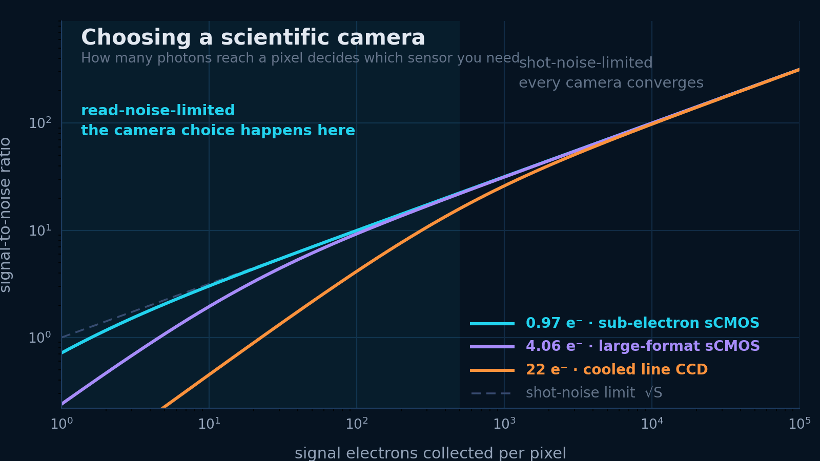 Scientific cameras
Choosing a scientific camera
Which sensor technology your photon budget actually needs, why rolling and global shutter answer different questions, and how to check the data can leave the camera at all.
Open guide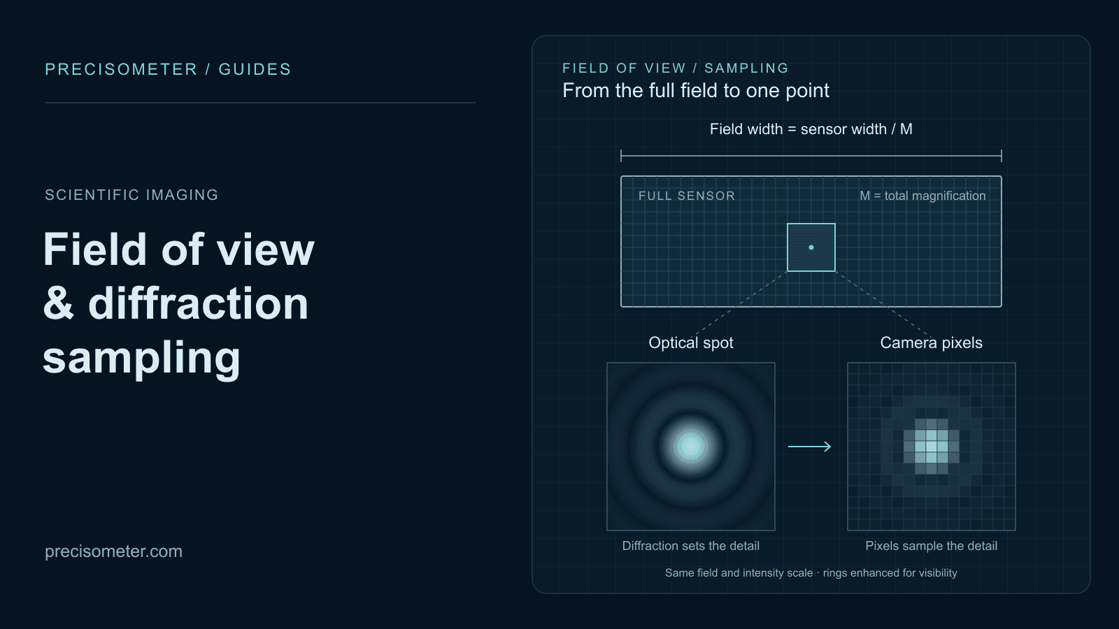 Scientific imaging
Calculating camera field of view and diffraction sampling
Calculate object-space pixel size, field of view, Rayleigh resolution, and sampling using real MAX and sMAX sensor formats.
Open guideSpecify the system
Tell us the specimen size and the clearing protocol.
Those two facts fix the sheet geometry, the objective, and the sensor format between them.