Imaging calculator
Calculating camera field of view and diffraction sampling
Connect sensor size, pixel pitch, magnification, wavelength, and numerical aperture—then see whether the camera captures the field and samples the optical detail correctly.
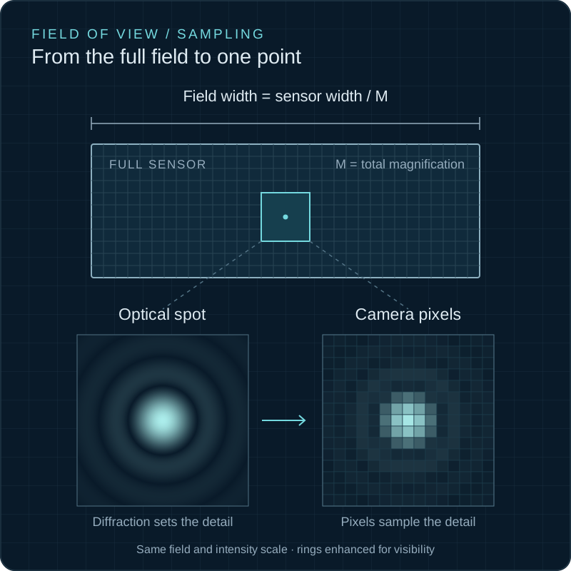
Field of view
Sensor dimensions divided by total magnification.
Object-space pixel
Camera pixel pitch divided by total magnification.
Diffraction sampling
Compare object-space pixels with 0.61λ/NA.
Two independent questions
Will the specimen fit, and will its detail be sampled?
A large sensor increases the specimen area recorded in one exposure. Small pixels increase sampling density. Neither automatically improves optical resolution: the objective, wavelength, numerical aperture, tube lens, relay optics, and camera port determine what information reaches the detector.
Calculate field of view and sampling separately. A system can have an excellent field of view but undersample diffraction-limited detail—or sample extremely finely while recording an unnecessarily small area and generating excessive data.
FOVx = sensor width ÷ magnification
Use physical sensor dimensions and the complete camera-port magnification.
Pixelobject = pixel pitch ÷ magnification
This is the sample distance represented by one camera pixel.
d = 0.61λ ÷ NA
A common lateral Rayleigh-resolution estimate for incoherent imaging.
Interactive FOV & sampling calculator
Use real MAX and sMAX sensor dimensions
Field of view
222 × 222 µm
Object-space pixel
108 nm
Rayleigh scale
280 nm
Pixels / Rayleigh
2.58
sMAX04BM
13.31 × 13.31 mm · 2048 × 2048 · 6.5 µm pixels
The Rayleigh scale is 0.61λ/NA. “Well sampled” here means roughly 2–3.5 pixels across that scale. Confirm the objective field number, camera-port optics, aberrations, and usable image circle before final selection.
View cameraInterpret the result
Undersampling loses information; extreme oversampling dilutes photons and throughput
Undersampled
<2 pixels per Rayleigh scale
Fine optical structure may alias or disappear. Increase magnification, use smaller pixels, or accept lower resolved detail.
Well sampled
About 2–3.5 pixels
A practical range for preserving diffraction-limited information without excessive data or photon spreading.
Oversampled
>3.5 pixels
The image may be smooth, but additional pixels do not create new optical resolution. Consider lower magnification or binning.
Before final selection
Check the complete optical interface
- Confirm the camera port’s actual relay magnification—not only the objective label.
- Verify that the microscope image circle covers the sensor without vignetting.
- Use emission wavelength for fluorescence sampling calculations.
- Include binning, cropping, and any intermediate magnification optics.
- Check objective field number, tube-lens compatibility, and aberration correction.
- Balance sampling against QE, read noise, frame rate, and data volume.
Choose the complete camera
Sampling is one part of scientific-camera selection.
Continue with QE and read-noise comparison, or browse the complete scientific-camera range.