Imaging comparison
Choosing a live-cell imaging system
Start with the number of culture vessels, the positions you need to revisit and the biological signal you want to follow. A single-position microscope, a motorised imaging system and a multilayer culture monitor solve different problems. The comparison below separates those workflows before you choose magnification or fluorescence.
Discuss your experiment →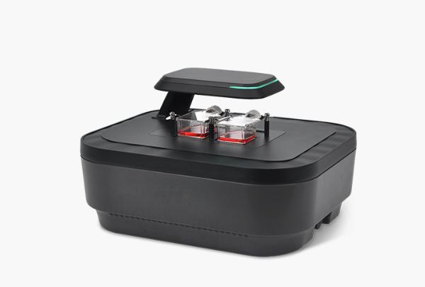
Compare the instruments
Specifications from the product datasheets. Open an instrument for the complete configuration, limitations and download. On a narrow screen, scroll the table horizontally.
| Instrument / workflow | Objectives | Imaging mode | Positions / vessels |
|---|---|---|---|
| Live-cell imaging system with interchangeable objectives → | 2×, 4× and 10×; user-interchangeable | Brightfield and two fluorescence channels | Motorised XYZ; multiple positions; autofocus and manual focus |
| Compact live-cell imaging system → | 2×, 4× or 10× brightfield; 4× or 10× fluorescence | Brightfield; brightfield with green or red fluorescence | Motorised XYZ, multiple positions; manual and autofocus |
| Dual-vessel live-cell imaging system → | 4× / 10× / 20×, interchangeable | Brightfield and two fluorescence channels | Up to two vessels; holder-dependent |
| Six-plate live-cell imaging system → | 4× / 10× / 20×, interchangeable | Brightfield and two fluorescence channels | Up to six vessels; holder-dependent |
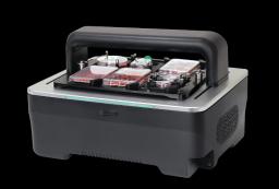
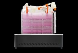
| Multilayer culture monitoring system → | 2× | Brightfield | Multilayer chambers up to ten layers; images acquired from bottom layer |
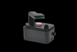
| Benchtop digital microscope → | 2×, 4× or 10×; configuration-dependent | Brightfield; optional green or red fluorescence | Single |
The multilayer system images the bottom layer only. The benchtop microscope has a single imaging position. Vessel capacity and fluorescence depend on the selected configuration and holder.
Choose the vessel workflow first
For repeated observations of one field, the benchtop digital microscope offers a single imaging position. For several fields or wells, choose a motorised multiposition system. The compact imager is available with different objective and fluorescence configurations; the interchangeable-objective system lets you change between 2×, 4× and 10×. Neither description should be read as a guarantee that an arbitrary plate or holder will fit.
The dual-vessel and six-vessel systems are relevant when you need independent cultures in the same experiment. Capacity is holder-dependent: send the vessel manufacturer, format and dimensions before treating “two” or “six” as a confirmed configuration. More positions, z-planes and channels also lengthen an acquisition cycle. Confirm that the complete schedule fits your required time interval.
For culture expansion in multilayer vessels, the dedicated monitor images the bottom layer of vessels with up to ten layers. It does not provide images of every layer. Use bottom-layer observations as a defined sampling strategy, and establish whether they represent the rest of your culture.
Match field of view and fluorescence to the readout
Low magnification gives context for growth and confluence; higher magnification can reveal smaller structures but covers less culture area per field. For example, the dual-vessel and six-vessel systems list fields of view of 2.0 × 1.5 mm at 4× and 0.5 × 0.4 mm at 20×. Check whether you need representative fields or stitched coverage. Objective magnification alone does not establish optical resolution.
Brightfield can support morphology, confluence and wound-area measurements without a fluorescent label. Fluorescence adds a labelled readout, but “green” or “red” is not enough to confirm dye compatibility. Send the fluorophore and excitation/emission requirements, then compare them with the exact filter set. Compact and benchtop variants use configuration-specific channels; do not assume they have the two-channel configuration of the larger systems.
For a long experiment, evaluate exposure, acquisition interval and focus on the actual cells. A software feature such as autofocus, stitching or cell counting needs a sample-specific check. A fluorescence count derived from images is also a different measurement from the concentration of a suspension measured in a counting chamber.
Check the complete incubator installation
Measure usable shelf width, depth and height, door clearance and cable access. Include the holder, loaded vessel and any cable bend rather than comparing the instrument body alone. The listed recommendation of an incubator above 170 L for the dual-vessel system or above 230 L for the six-vessel system is only an initial screen; usable clearance must still be checked.
Confirm the operating environment, cleaning method, external computer and data exports with the chosen configuration. Ask for an example image and exported result relevant to your assay. Before ordering, establish which holder, objectives, fluorescence options, software functions and computer are included, and which installation, training and service arrangements are available.
Instruments for this workflow
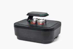
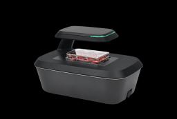
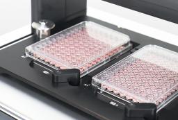
Check the exact configuration and assay compatibility on the product page. Inclusion does not establish validation for every sample.
Sources and scope
The comparison uses specifications in the linked product datasheets. This is selection guidance, not a claim of performance validation for your sample. For research use.
Details for instrument selection
- Cell type, assay and required readout
- Exact vessel format, number of vessels and imaging positions
- Magnification, fluorophores, interval and experiment duration
- Incubator model, usable internal dimensions and cable access
Discuss your experiment
Tell us what you need to measure. Next, add your contact details and review the enquiry before sending.
Selection help and applications
Cell-counting guide
Choosing a cell counter for cell culture →
Choose a counting method by cell type, concentration, viability stain and sample workload. Compare counters and plan a sample-specific evaluation.
Application example
Tracking wound closure with time-lapse imaging →
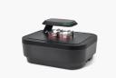
A documented NIH-3T3 scratch-assay example: brightfield imaging every 30 minutes for 72 hours, wound-area analysis and interpretation limits.
Application example
Following a cytotoxic response over time →
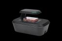
A documented HeLa-cell example combining brightfield confluence and green fluorescence during a 48-hour nocodazole exposure.