Cell-counting guide
Choosing a cell counter for cell culture
Choose a counting method around the suspension you actually measure: cell size, concentration, aggregates, debris and the reason you need a count. Routine seeding, viability monitoring and somatic-cell counting in milk are different workflows. A useful selection compares the measurement and sample preparation as well as the instrument.
Discuss your experiment →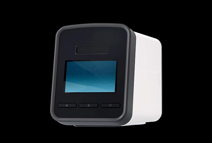
Compare the instruments
Specifications from the product datasheets. Open an instrument for the complete configuration, limitations and download. On a narrow screen, scroll the table horizontally.
| Instrument | Cell diameter | Concentration range | Loading volume |
|---|---|---|---|
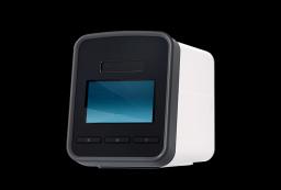
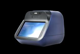
| Automated brightfield cell counter → | 5–60 µm | 1 × 10⁴ to 1 × 10⁷ cells/mL | 20 µL per chamber |
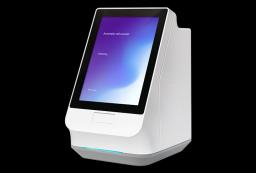
| Four-channel automated cell counter → | 5–60 µm | 1 × 10⁴ to 2 × 10⁷ cells/mL | 20 µL/channel |
Define the sample and the quantity to report
Record the cell type, expected diameter distribution and concentration before choosing a preset. Both automated cell-culture counters below list a cell-diameter range of 5–60 µm. That range is a catalogue specification, not evidence that every cell type or mixed suspension within it is measured equally well. Review the image overlays for small cells, debris, touching cells and aggregates.
Total concentration, viable concentration and percentage viability are different results. The four-channel counter lists trypan blue or Erythrosin B for viability assessment. Confirm the stain, mixing ratio and incubation/read timing for your method. Membrane-integrity staining measures a particular aspect of viability; it does not establish every aspect of cell function.
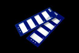
For milk, use the separate fluorescence-based somatic-cell workflow and its slide/stain kit. Its sample matrix and analysis are different from routine mammalian cell-culture counts. Do not substitute it for a culture counter solely because both report cells per millilitre.
Compare the working range and the analysed volume
The brightfield counter lists 1 × 10⁴ to 1 × 10⁷ cells/mL. Its quick, normal and precise modes list analysed volumes of 0.15, 0.9 and 3.6 µL, with approximate processing times of 20, 30 and 100 seconds per test. Choose the mode around the required measurement, not just the shortest processing time. These are instrument processing figures; preparation and result review add time.
The four-channel system lists 1 × 10⁴ to 2 × 10⁷ cells/mL, with a recommended range of 1 × 10⁵ to 1 × 10⁷ cells/mL. It loads 20 µL per channel and lists approximately 15 seconds per channel. Four channels mean up to four samples on a slide; they do not mean four fluorescence colours or four simultaneous measurements completed in 15 seconds.
Loading volume is not the same as analysed volume. If cells are dilute, fewer counted objects can make an estimate more sensitive to sampling variation. If a sample is too concentrated, dilution may be needed to avoid overlapping objects. Record the dilution factor and check whether the instrument applies it automatically before correcting the result again.
Evaluate with your own samples
Begin with representative samples and predefined acceptance criteria. Mix consistently, prepare independent chamber loads and repeat measurements across the concentrations and viability levels relevant to your work. Review saved images alongside the numeric result so a change in segmentation is not mistaken for a biological difference.
A dilution series can test proportionality; independent loads can test repeatability. Agreement with a manual chamber is useful context, but that chamber also has sampling and operator uncertainty. Neither agreement alone nor a high correlation establishes absolute accuracy. Record the sample preparation, stain, preset, analysis gates and dilution calculation with each comparison.
For occasional counts or visual checks, a disposable haemocytometer may suit the workflow, provided the microscope and operator method are appropriate. For routine batches, consider how many samples arrive together, slide compatibility, preparation, image review and export. Confirm consumable supply and software functions before selecting by nominal seconds per test.
Instruments for this workflow
Check the exact configuration and assay compatibility on the product page. Inclusion does not establish validation for every sample.
Sources and scope
The comparison uses specifications in the linked product datasheets. This is selection guidance, not a claim of performance validation for your sample. For research use.
Methodological background for evaluating counting methods: NIST — cell-counting precision and proportionality. This reference does not imply endorsement of the instruments offered.
Details for instrument selection
- Cell type, diameter and expected concentration
- Total count, viable concentration or viability percentage
- Stain, dilution method and samples per batch
- Required image review, export and compatible consumables
Discuss your experiment
Tell us what you need to measure. Next, add your contact details and review the enquiry before sending.
Selection help and applications
Imaging comparison
Choosing a live-cell imaging system →
Compare six imaging systems by vessel workflow, objectives, fluorescence and incubator fit, then specify the configuration for your experiment.
Application example
Tracking wound closure with time-lapse imaging →
A documented NIH-3T3 scratch-assay example: brightfield imaging every 30 minutes for 72 hours, wound-area analysis and interpretation limits.
Application example
Following a cytotoxic response over time →
A documented HeLa-cell example combining brightfield confluence and green fluorescence during a 48-hour nocodazole exposure.