Application example
Tracking wound closure with time-lapse imaging
A scratch assay follows the changing cell-free area in a cultured monolayer. In this documented experiment, repeated brightfield images were used to compare NIH-3T3 fibroblasts under different serum and drug conditions. The useful output is the area trajectory together with the images that explain it.
Discuss your experiment →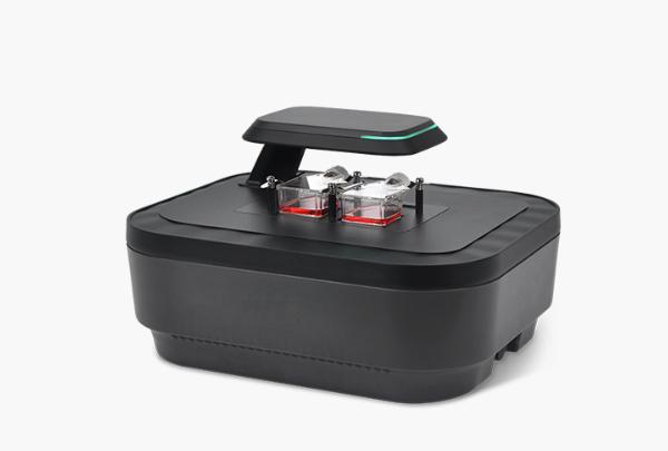
Reported experimental setup
- Cells and preparation
- NIH-3T3 fibroblasts; scratch at approximately 90% confluence; two PBS washes after scratching.
- Culture and comparison
- DMEM; 0%, 5% or 10% FBS; culture at 37 °C. Separate drug comparisons used 10% FBS.
- Acquisition
- Brightfield images every 30 minutes for 72 hours.
- Readout
- Segmented wound area at each time point; closure (%) = 100 × [A(0) − A(t)] / A(0).
- 1
Prepare matched monolayers and a defined scratch
- 2
Revisit the same fields on a fixed schedule
- 3
Inspect segmentation and compare area trajectories
What was reported
The application note reports faster closure with 10% FBS than with 5% or 0% FBS. Drug-treated cultures produced different closure patterns. Inspecting the accompanying images helped distinguish changes in morphology and cell coverage that a final endpoint alone would not explain.
Interpretation limits
Wound closure is influenced by migration, proliferation, cell spreading and cell death. It is not a migration-specific measurement by itself. Initial wound geometry, field selection and segmentation settings need control. The source does not provide raw data or a complete replicate-level statistical dataset here; no numerical effect size or accuracy claim is inferred.
Translate the example to your assay
Use an initial image to define the wound area, keep the same field registered over time and review the segmentation overlay at early and late time points. Retain representative images alongside the area values. Include untreated or vehicle controls appropriate to your assay; additional readouts may be needed to separate migration from growth or cell death.
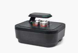
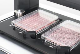
A motorised multiposition brightfield system is a relevant starting point when several fields or conditions must be revisited in an incubator. Confirm the vessel holder, complete acquisition-cycle duration and availability of the required wound-area analysis function in the proposed software configuration. The settings above describe this experiment, not a universal protocol.
Instruments for this workflow
Check the exact configuration and assay compatibility on the product page. Inclusion does not establish validation for every sample.
Sources and scope
Original summary of an instrument application note on wound-closure dynamics. Reported experimental example, not an independent validation or work performed by Precisometer. No experimental images or curves from that note are reproduced here.
Details for instrument selection
- Cell type, vessel and scratch method
- Number of fields, conditions, interval and duration
- Required wound-area output and controls
Discuss your experiment
Tell us what you need to measure. Next, add your contact details and review the enquiry before sending.
Selection help and applications
Imaging comparison
Choosing a live-cell imaging system →
Compare six imaging systems by vessel workflow, objectives, fluorescence and incubator fit, then specify the configuration for your experiment.
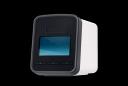
Cell-counting guide
Choosing a cell counter for cell culture →
Choose a counting method by cell type, concentration, viability stain and sample workload. Compare counters and plan a sample-specific evaluation.
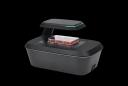
Application example
Following a cytotoxic response over time →
A documented HeLa-cell example combining brightfield confluence and green fluorescence during a 48-hour nocodazole exposure.