Compact live-cell imaging system
Incubator-compatible time-lapse imaging with autofocus, z-stacking and brightfield or fluorescence configurations.
Technical summary with specifications and configuration choices.
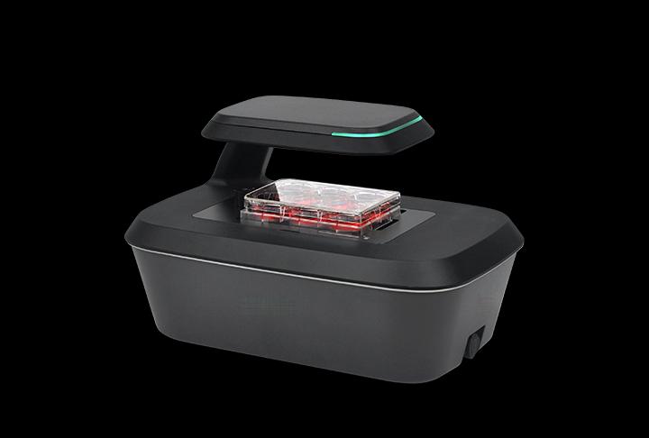
Features
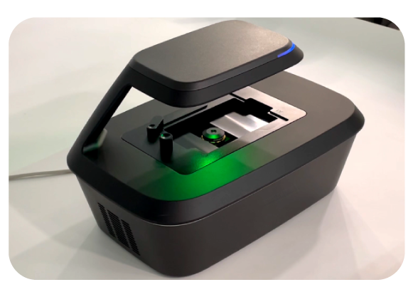
- Incubator imaging with a fixed culture vessel and a moving camera.
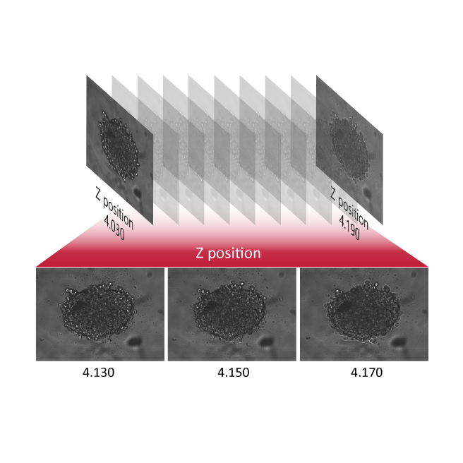
- Automated multiple-position time-lapse acquisition, autofocus and z-stacking.
- Brightfield models and models with a green or red fluorescence channel.
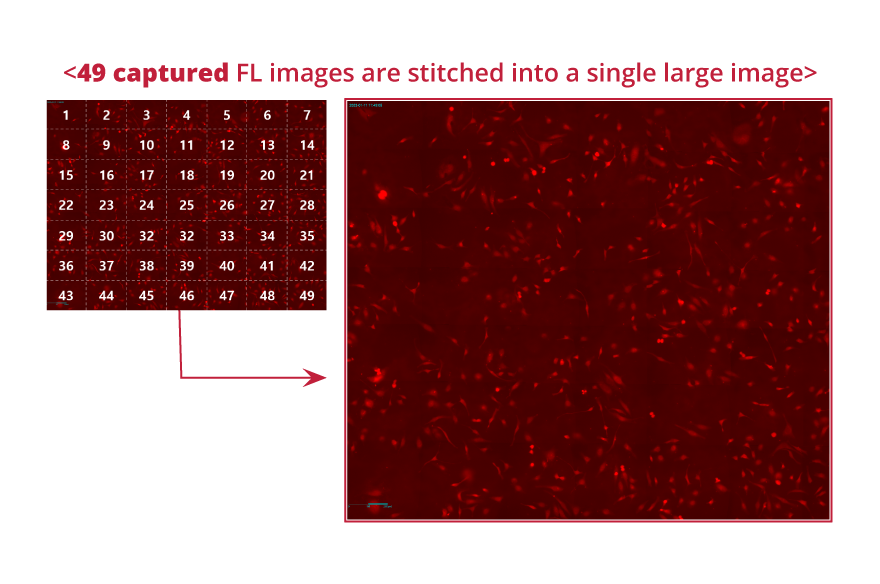
- Analysis tools support stitching, confluence and time-lapse review.
Images and workflow examples
Select an image to view it at full size.
Technical specifications
| Imaging configurations | Brightfield; brightfield with green or red fluorescence |
|---|---|
| Objective configurations | 2×, 4× or 10× brightfield; 4× or 10× fluorescence |
| Fluorescence options | Green Ex 470/40 nm, Em 510 nm long-pass; red Ex 525/30 nm, Em 570 nm long-pass |
| Camera / illumination | 5 MP CMOS / LED |
| Fields of view | 2× 2.0 × 1.5 mm; 4× 1.4 × 1.0 mm; 10× 0.7 × 0.5 mm |
| Motion and focus | Motorised XYZ, multiple positions; manual and autofocus |
| Culture vessels | Plates up to 96 wells, flasks, dishes and slides; holder-dependent |
| Exports | TIFF, AVI, CSV; JPEG and PNG |
| Computer | Windows 10 or later |
| Dimensions | 226 × 358 × 215 mm |
| Weight | 5.6 kg |
| Operating environment | 10–40 °C; 20–95% relative humidity |
| Power input | 100–240 V AC, 50/60 Hz |
Applications
- Monitor cell growth and confluence during culture.
- Compare treated and control cultures in time-lapse experiments.
Configurations and accessories
- Choose objective magnification and brightfield, green or red fluorescence before ordering.
- Select the holder for your plate, flask, dish or microscope slide.
- Include the incubator model, internal dimensions and cable access in your enquiry.
Selection notes
- For research use. Match the objective, vessel holder and fluorescence configuration to your assay when requesting a quotation.
Selection help and applications
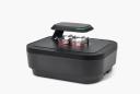
Imaging comparison
Choosing a live-cell imaging system →
Compare six imaging systems by vessel workflow, objectives, fluorescence and incubator fit, then specify the configuration for your experiment.
Application example
Tracking wound closure with time-lapse imaging →
A documented NIH-3T3 scratch-assay example: brightfield imaging every 30 minutes for 72 hours, wound-area analysis and interpretation limits.
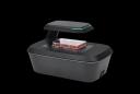
Application example
Following a cytotoxic response over time →
A documented HeLa-cell example combining brightfield confluence and green fluorescence during a 48-hour nocodazole exposure.
Discuss your experiment
Tell us what you need to measure. Next, add your contact details and review the enquiry before sending.