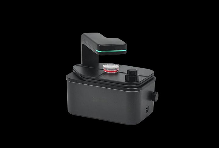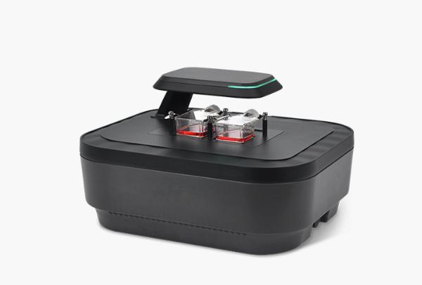
Live-cell imaging system with interchangeable objectives
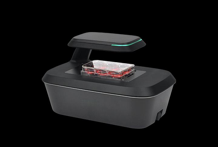
Automated multiposition time-lapse imaging in brightfield and two fluorescence channels, with interchangeable 2×, 4× and 10× objectives.
Specifications and datasheet →Compare single-position microscopes, automated live-cell imaging systems, two-vessel and six-vessel instruments, and multilayer culture monitors. Choose by vessel capacity, field of view and fluorescence requirements.
Automated multiposition time-lapse imaging in brightfield and two fluorescence channels, with interchangeable 2×, 4× and 10× objectives.
Specifications and datasheet →Incubator-compatible time-lapse imaging with autofocus, z-stacking and brightfield or fluorescence configurations.
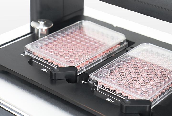
Specifications and datasheet →Monitor two culture vessels in parallel inside an incubator using brightfield and two fluorescence channels.
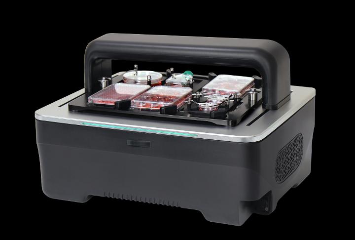
Specifications and datasheet →Time-lapse imaging for up to six culture plates in an incubator, with brightfield and two fluorescence channels.
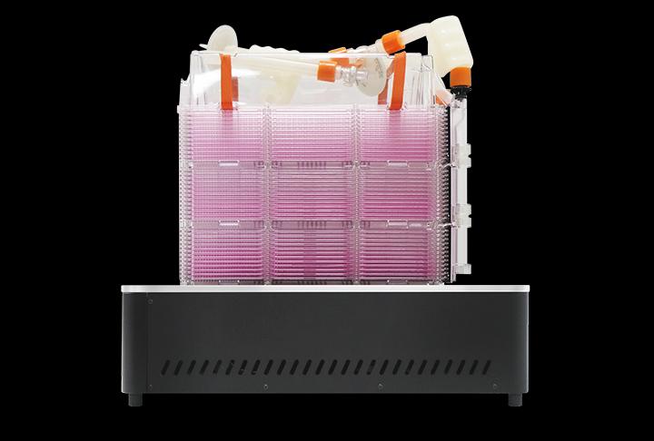
Specifications and datasheet →Use brightfield time-lapse imaging to monitor the bottom layer of a culture vessel with up to ten layers.
Specifications and datasheet →Compact microscope for observing cells, with brightfield and fluorescence configurations.
Specifications and datasheet →
Imaging comparison
Compare six imaging systems by vessel workflow, objectives, fluorescence and incubator fit, then specify the configuration for your experiment.
Application example
A documented NIH-3T3 scratch-assay example: brightfield imaging every 30 minutes for 72 hours, wound-area analysis and interpretation limits.
Application example
A documented HeLa-cell example combining brightfield confluence and green fluorescence during a 48-hour nocodazole exposure.
Tell us what you need to measure. Next, add your contact details and review the enquiry before sending.