Multilayer culture monitoring system
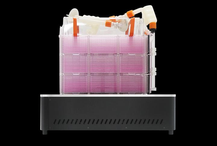
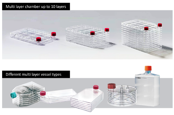
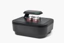
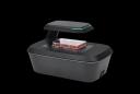
Use brightfield time-lapse imaging to monitor the bottom layer of a culture vessel with up to ten layers.
Technical summary with specifications and configuration choices.
Features
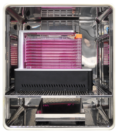
- Observe cells at the bottom layer while a multilayer vessel remains in the incubator.
- Schedule multiple imaging positions and assemble time-lapse videos.
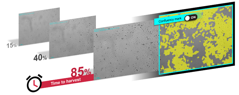
- Use confluence and growth-curve analysis to support harvest timing.
Images and workflow examples
Select an image to view it at full size.
Technical specifications
| Vessels | Multilayer chambers up to ten layers; images acquired from bottom layer |
|---|---|
| Imaging mode | Brightfield |
| Objective | 2× |
| Camera | 5 MP CMOS, 2592 × 1944 pixels |
| Stage | Motorised XYZ |
| Focus | Autofocus and manual |
| Imaging positions | Multiple |
| Analysis | Time-lapse, movie creation, confluence and growth curves |
| Exports | TIFF, AVI |
| Dimensions (H × W × D) | 132 × 330 × 450 mm |
| Weight | 11.6 kg |
| Operating environment | 10–40 °C; 20–95% relative humidity |
Applications
- Track growth during expansion in multilayer culture vessels.
- Inspect culture development without repeatedly moving vessels outside the incubator.
Configurations and accessories
- Specify the exact multilayer vessel and its dimensions for holder selection.
- Alternative holders for multilayer dishes and flasks can be assessed on request.
Selection notes
- For research use. Match the objective, vessel holder and fluorescence configuration to your assay when requesting a quotation.
Selection help and applications
Imaging comparison
Choosing a live-cell imaging system →
Compare six imaging systems by vessel workflow, objectives, fluorescence and incubator fit, then specify the configuration for your experiment.
Application example
Tracking wound closure with time-lapse imaging →
A documented NIH-3T3 scratch-assay example: brightfield imaging every 30 minutes for 72 hours, wound-area analysis and interpretation limits.
Application example
Following a cytotoxic response over time →
A documented HeLa-cell example combining brightfield confluence and green fluorescence during a 48-hour nocodazole exposure.
Discuss your experiment
Tell us what you need to measure. Next, add your contact details and review the enquiry before sending.