Integrated optoelectronic test system
RTS2-Omni-Imager
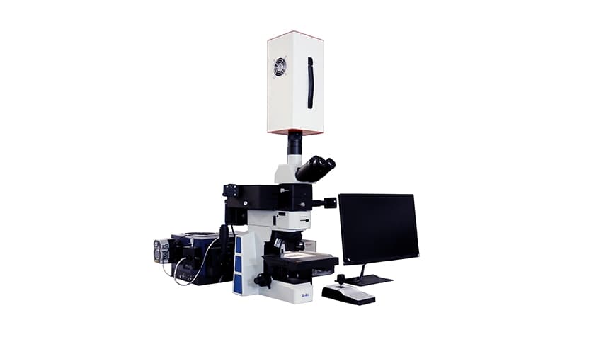
A Raman–hyperspectral combined microscopic spectroscopy system uniting dark-field scattering, steady-state and transient fluorescence, and Raman spectroscopy on one open, modular platform.
400–1000 nm
Spectral range
2.8 nm
Resolution
9 s
Per data cube
01 · Overview
One platform, four measurement modes
The modular RTS2-Omni-Imager moves beyond single-technique spectrometers. It records image and spectral information together for fast, in-situ, correlative analysis of physical, chemical, and biological processes at the nanoscale.
Push-broom hyperspectral imaging
Capture a complete data cube in roughly 10 seconds to one minute, depending on camera settings, without point-by-point scanning.
Patented focal-plane scanning
An internal precision motorized stage handles acquisition, focus, and depth-of-field adjustment through patented focal-plane scanning technology.
Large-area automated mapping
An optional motorized XY stage maps large samples and automatically stitches and corrects each swath into a seamless image.
Depth-resolved imaging
Motorized autofocus supports in-situ imaging across sample layers for large-area, multi-dimensional, and tomographic measurements.
02 · Technology
Dark-field microscopy meets hyperspectral imaging
Dark-field microscopy
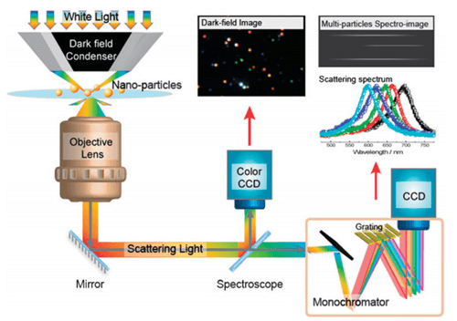
A dedicated illumination and observation geometry directs only sample-scattered light into the objective. Small particles, fibres, and interfaces stand out against a dark background, including structures below the conventional bright-field diffraction contrast limit.
Hyperspectral imaging
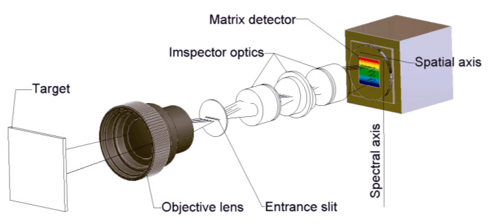
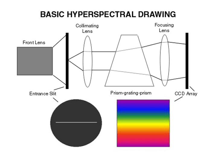
Tens to hundreds of continuous narrow spectral bands create a complete spectrum for every image point. A push-broom imaging spectrometer records one spatial line at a time, combining spatial and spectral axes into a calibrated three-dimensional data cube.
03 · System configuration
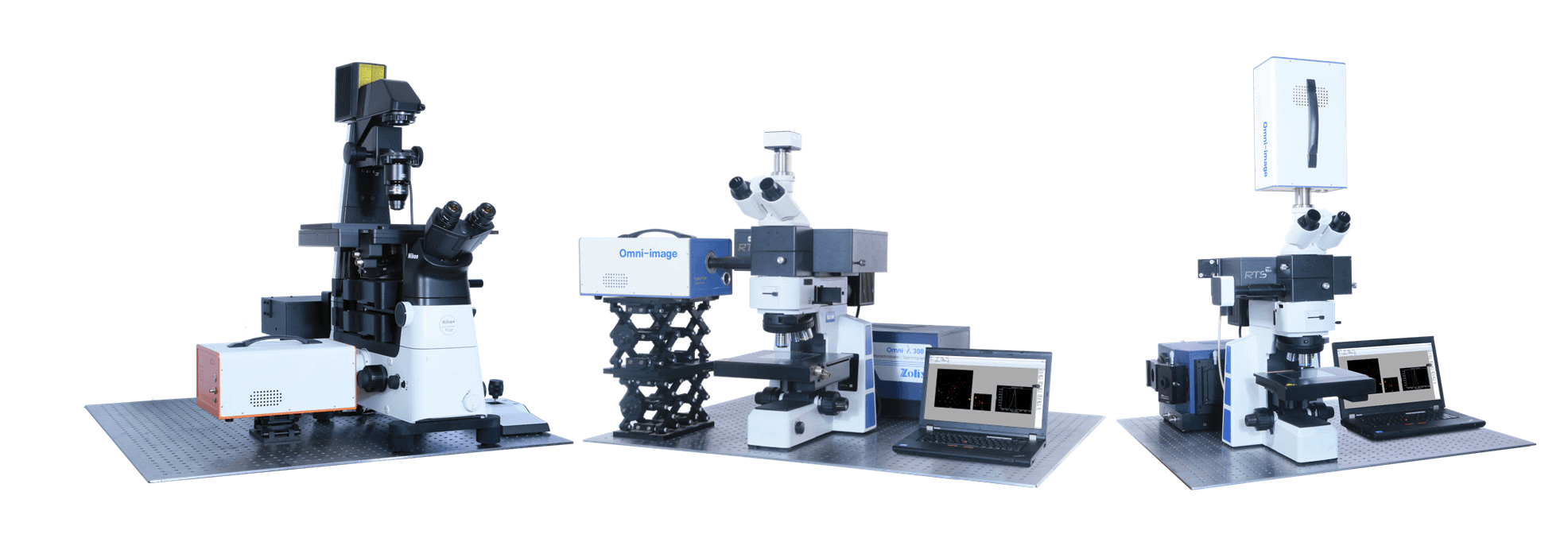
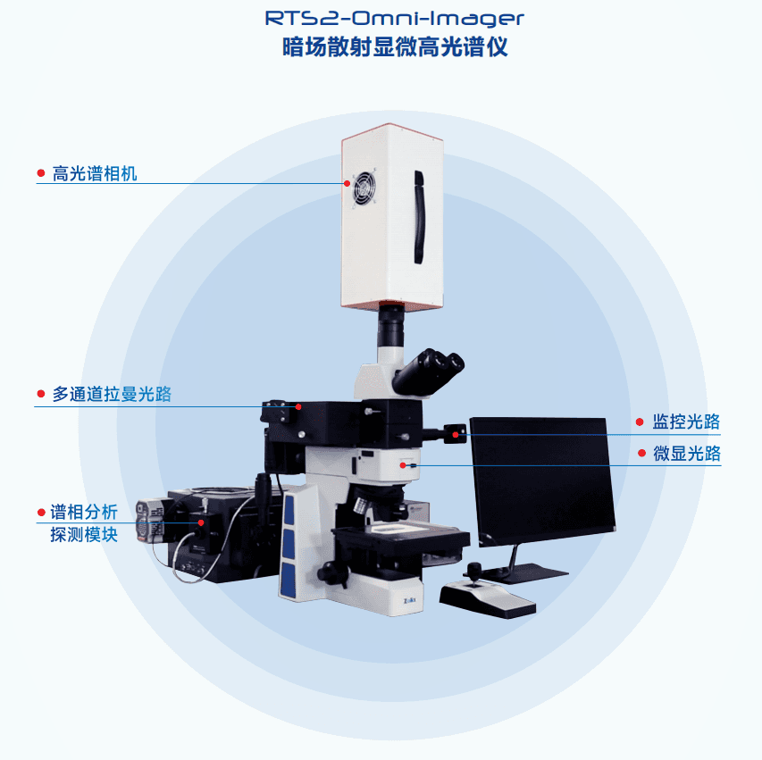
A modular optical architecture
A dark-field microscope, imaging spectrograph, hyperspectral camera, and high-sensitivity detector form the core. Optional laser and confocal paths add fluorescence and Raman excitation while keeping conventional spectrograph access available.
04 · Software & database
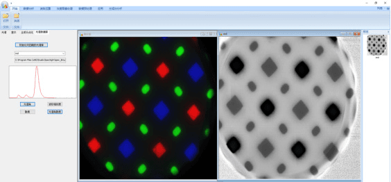
Modular spectral-image processing
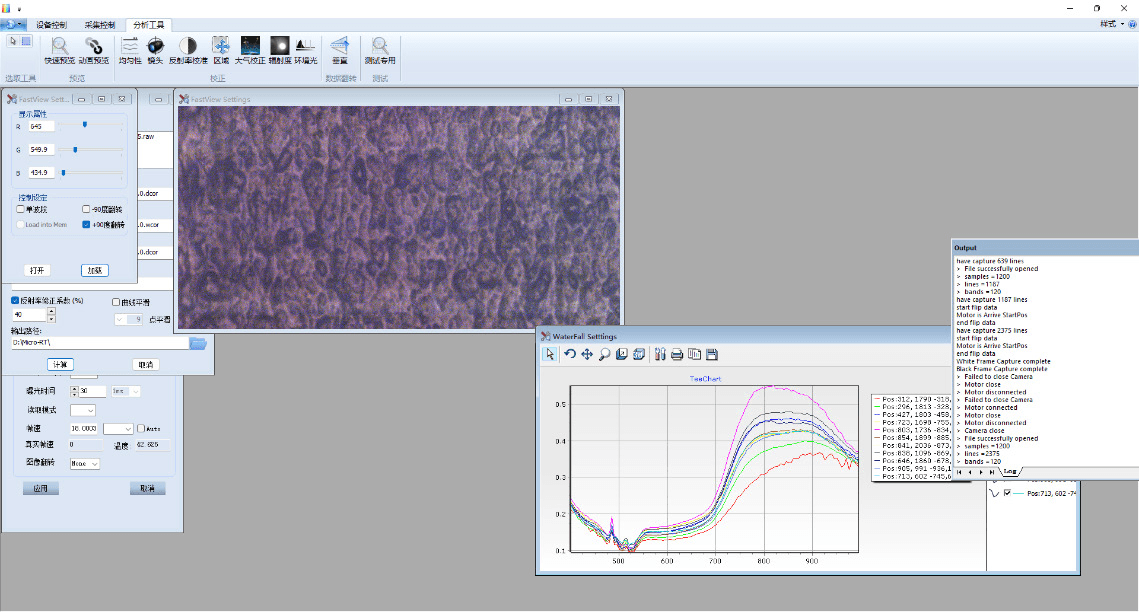
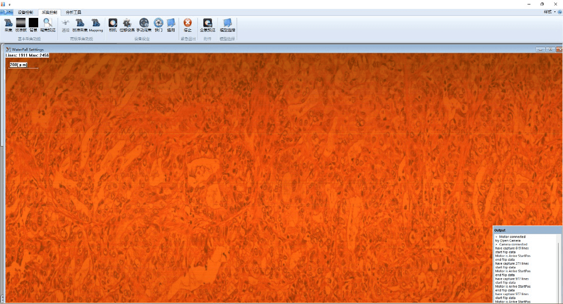
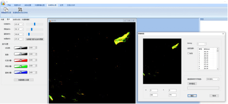
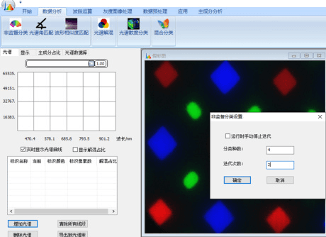
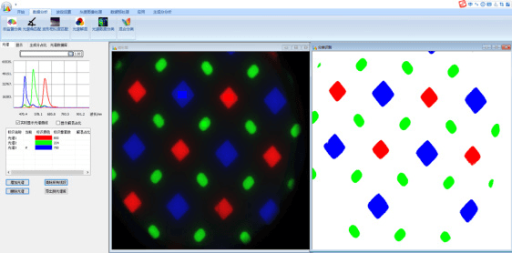
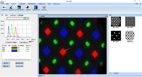
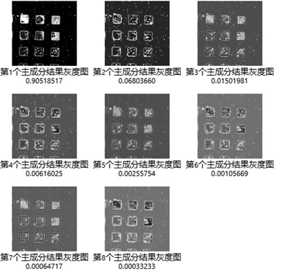
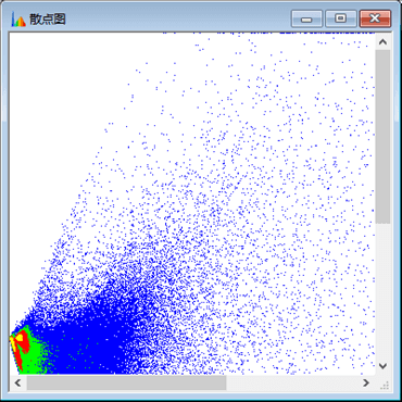
Image-processing and spectral-identification modules cover classification, spectral-angle matching, linear unmixing, divergence analysis, band math, PCA, calibration, retrieval, and spectral-database management.
Band extraction and frame selection
Select the wavelength range and channel count of interest, increasing acquisition speed when fewer bands are required.
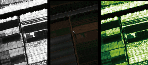
Calibration and retrieval
One-click reflectance calibration, batch processing, binning, cropping, format conversion, and model-based real-time retrieval.
05 · Applications & measured data
From single nanoparticles to perovskite films
Correlative image and spectrum acquisition supports dark-field scattering, nanomaterials research, life sciences, camouflage detection, and pharmaceutical or medical analysis.
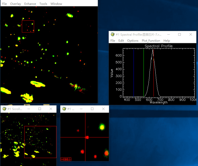
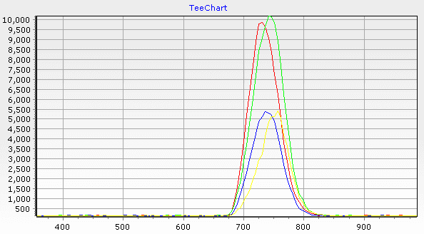
Nanoparticles in one field of view
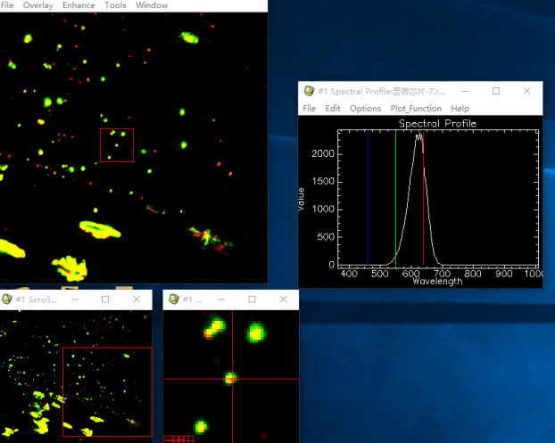
Polarization-resolved single gold nanorod
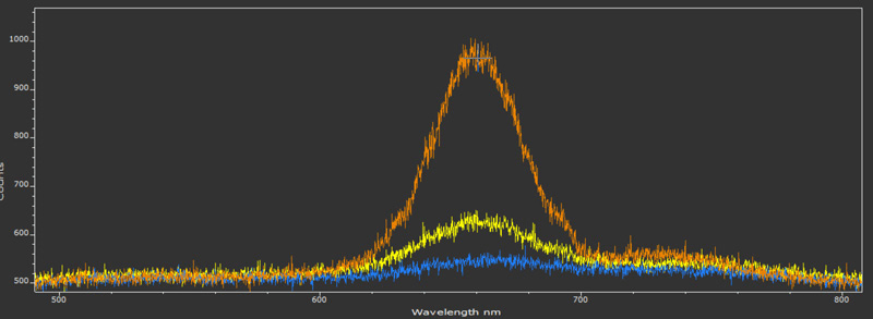
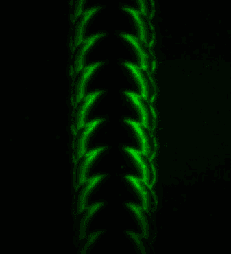
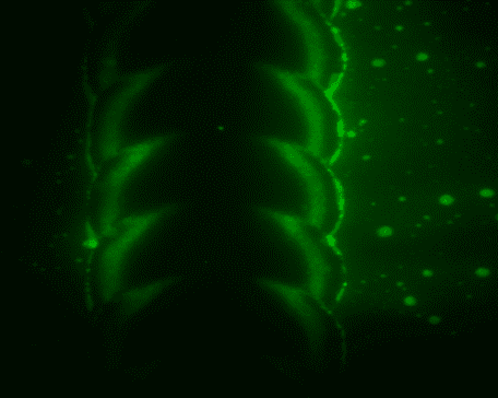
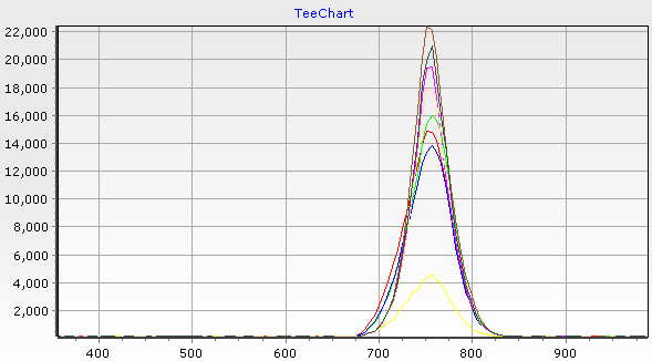
Perovskite fluorescence mapping
Excitation at 532 nm with detection around the 750 nm fluorescence peak.
06 · Technical specifications
Performance and configuration
Hyperspectral imaging
| Scanning method | Internal push-broom scanning |
|---|---|
| Focusing | Motorized, electronically controlled |
| Spectral range | 400–1000 nm |
| Spectral resolution | 2.8 nm |
| Spectral sampling | 0.6 nm |
| Detector | 2048 × 2048 cooled sCMOS; air- or water-cooled |
| Dynamic range | 16 bit |
| Image spatial resolution | 2048 × 1024 |
| Effective spectral channels | User-definable |
| Scan speed | 9 seconds per data cube |
Dark-field scattering & fluorescence
| Spectral range | 400–1000 nm |
|---|---|
| Spectral resolution | < 3 nm |
| Imaging speed | < 1 minute at 640 × 512 |
| Spatial resolution | < 2 µm |
Optional Raman module
| Raman spectral range | 90–5000 cm⁻¹ |
|---|---|
| Spectral resolution | ≤ 2.0 cm⁻¹ at 1800 g/mm |
| Silicon third-order peak SNR | > 30:1 |
Hardware configuration
| Microscope | Upright microscope with 100×, 50×, and 10× dark-field objectives; inverted microscope optional |
|---|---|
| Hyperspectral camera | Omni-Imager, F/2.4, 400–1000 nm |
| Control computer | All-in-one PC |
| Optional spectrograph | 320 mm or 500 mm focal length, three gratings, motorized shutter, and deep-cooled back-illuminated detector |
| Optional excitation | Raman lasers plus CW or pulsed fluorescence sources, including picosecond, femtosecond, and supercontinuum sources |
| Power supply | 220 V |
| Operating environment | 20 ± 5 °C; ≤ 85% RH; isolated from high-power electrical equipment and vibration sources |
Specifications are based on manufacturer documentation and may change with configuration. Precisometer will confirm the final optical, detector, excitation, software, and accessory package before quotation.
Local support
Configure the RTS2 around your experiment
We help define microscope geometry, spectral range, detector, Raman excitation, mapping stage, environmental accessories, acceptance criteria, installation, and training, with in-person support in the Netherlands and Belgium.