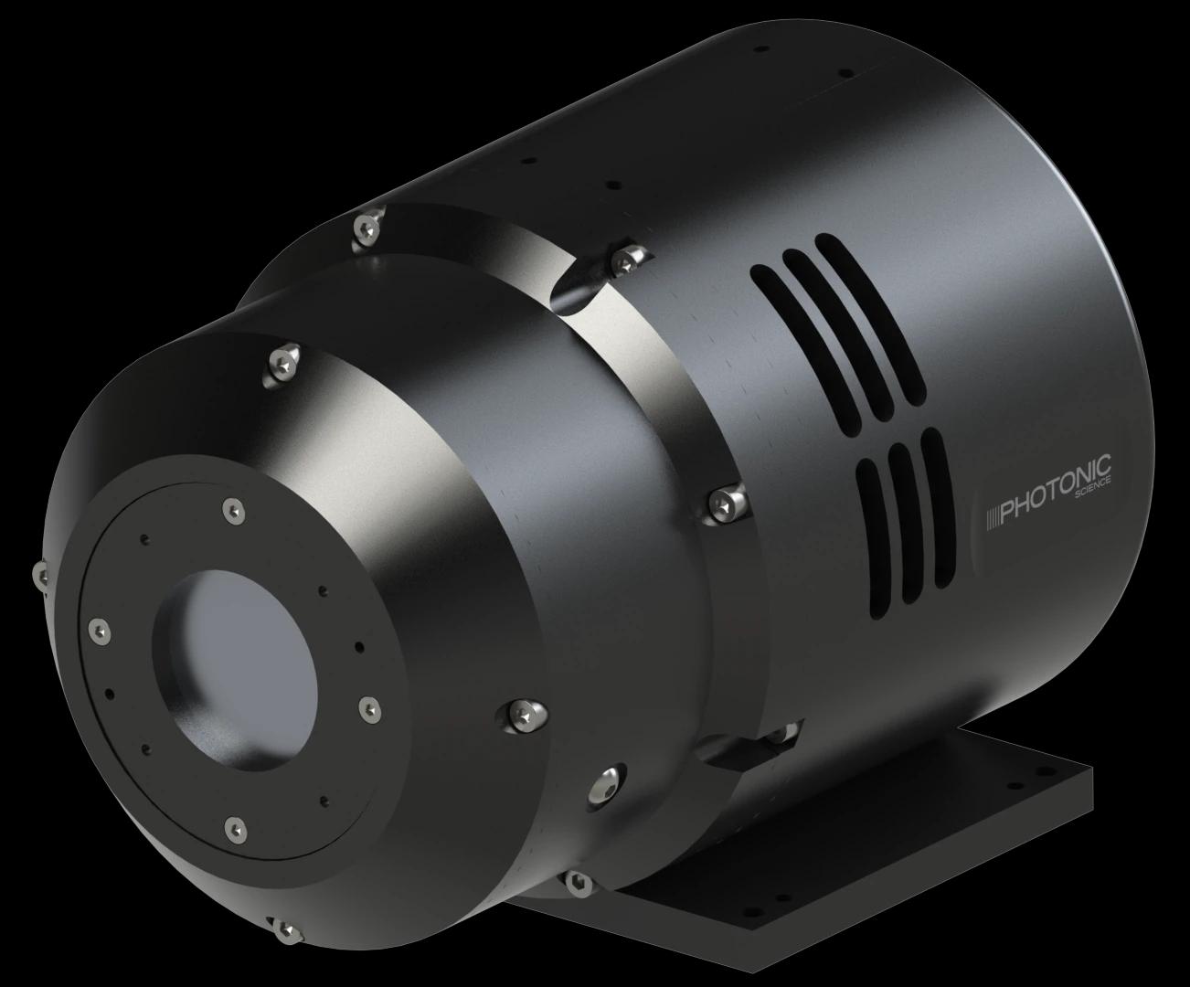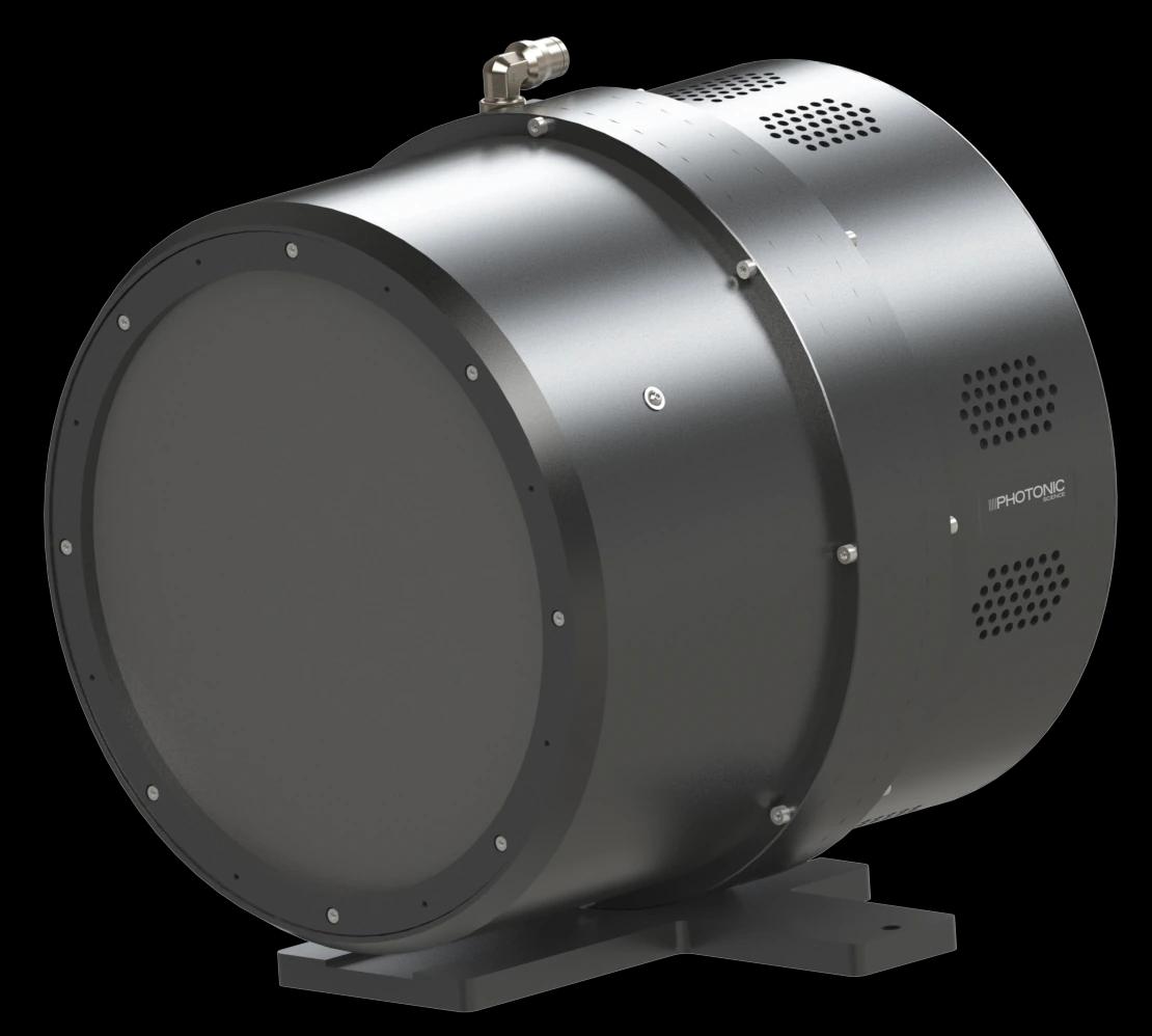
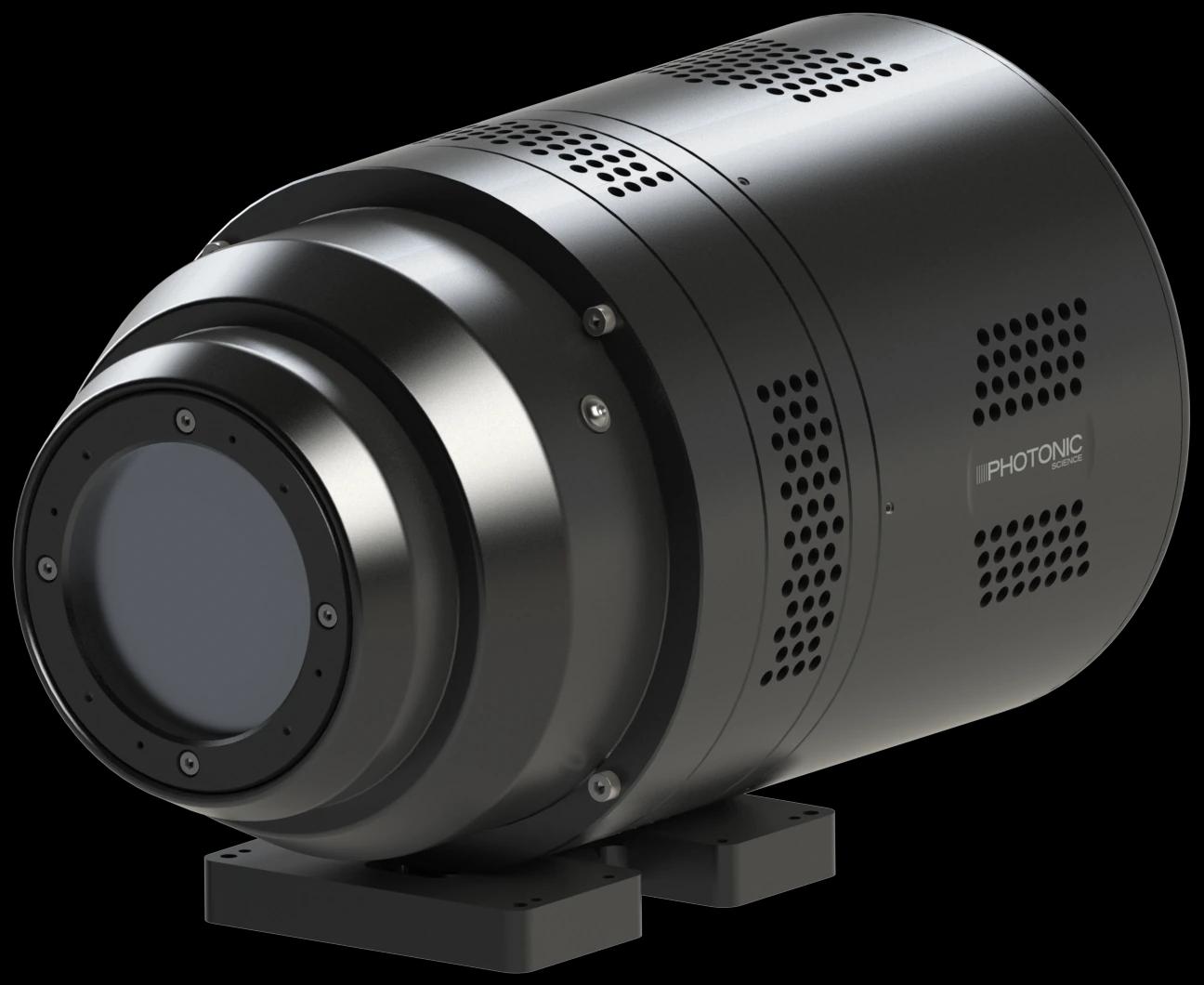
X-ray sCMOS 16MP Cooled
Fibre-optic coupled 4096 × 4096 X-ray detection with air or water cooling and two standard active input sizes.
- Resolution
- 4096 × 4096 pixels
- Input sizes
- 36.9 × 36.9 mm / 66.5 × 66.5 mm
Radiography systems
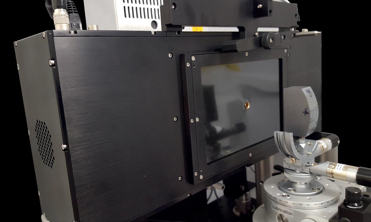
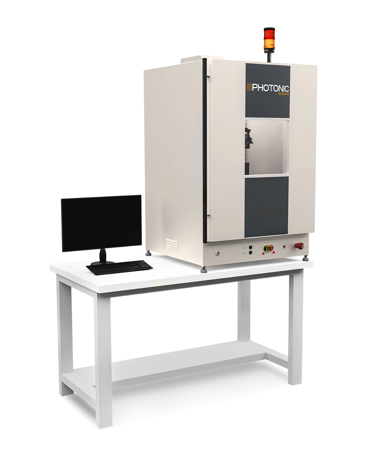
An integrated X-ray cabinet for short-exposure imaging using a 50 W minifocus source.
From photon to signal
No system-level QE or DQE curve is published for this cabinet. Detector efficiency depends on the configured camera, scintillator, X-ray spectrum and sample geometry; source and detector specifications are listed below.
The details behind the selection
Compare sensor configurations, coating options, operating limits and integration details directly below. Check the operating mode and temperature alongside each performance value.
| Parameter | High Resolution | Rapid Imaging |
|---|---|---|
| X-ray source | 60 kVp microfocus | 50 kVp minifocus; 50 W |
| Resolution at target | 3–5 µm | 30 µm |
| Magnification | 5–10× | 1:1 |
| Typical exposure in model feature list | Approximately 2 s; sample dependent | >1 s; sample dependent |
| Cabinet (W × D × H) | 694 × 782 × 1200 mm | 694 × 782 × 800 mm |
| Cabinet | Fully self-shielded benchtop cabinet | Fully self-shielded benchtop cabinet |
| Rapid-imaging input | Detector selected for the application | Up to 95 mm diagonal |
The introductory text describes rapid exposures below 1 s, while the model feature list gives >1 s typical. Both are retained with their context; exposure depends on sample, source setting and detector. Resolution also depends on magnification and scintillator.
| Key Specifications | High-Resolution System | Rapid Imaging System |
|---|---|---|
| Resolution at the target | 3 µm at 2.5W with 5x magnification 5 µm at 8W with 5x magnification* | ~30 µm |
| Magnification range | 5x to 10x | ~1:1 |
| Area of illumination target | 7.34 mm2 at 5x magnification 3.67 mm2 at 10x magnification | ~36.7 mm2 or ~65.5 mm2 |
| Typical exposure time (sample dependent) | ~2 sec | >1 sec |
| Detector Specifications* | High-Resolution System | Rapid Imaging System |
|---|---|---|
| Number of Pixels | 4096 x 4096 | 4096 x 4096 |
| FOV | 36.7 x 36.7 mm | 36.7 x 36.7 mm or 65.5 x 65.5 mm |
| Effective Pixel Size | 9 µm x 9 µm | 9 µm x 9 µm or 14.1 µm x 14.1 µm |
| Dynamic Range | 12000:1 with all corrections | |
| Frame Rate | 4.5 fps at full sensor resolution; 18 fps at half sensor resolution | |
| Full Well Capacity | 65000 to 70000 typical in mixed gain mode | |
| Read Out Noise | <5 electrons RMS at full sensor resolution and mixed gain mode <10 electrons RMS at half sensor resolution | |
| Dark Current | <1 electron/pixel/second air cooled with all corrections | |
| Digitisation | 16-bit | |
| Peak QE | 58% at scintillator emission wavelength (without microlens) | 58% at scintillator emission wavelength |
| Exposure | 50 microseconds up to 60 minutes | |
| Spatial Resolution | 30 lp/mm | 30 lp/mm or 20 lp/mm |
| Detector Interface | Gigabit Ethernet | |
| Energy Range | 5 keV - 55 keV with Gadox:Tb | |
| X-ray Source | High-Resolution System | Rapid Imaging System |
|---|---|---|
| Power | 8W | 50W |
| Spot Size | 5 µm at 8W | 50 µm at 50W |
| Energy Range | 20 - 60 kVp | |
| X-ray Beam Angle | 40º round beam | |
| Target Material | Tungsten | |
| Cooling Method | Air-cooled | |
Build your enquiry
Capture the system requirements and integration details you want to discuss with Precisometer.
Technical overview
An integrated X-ray cabinet for short-exposure imaging using a 50 W minifocus source.
| Source | 50 W minifocus |
|---|---|
| Object resolution | 30 µm |
| Acquisition | Exposures below 1 s; input up to 95 mm diagonal |
| Integration | sCMOS detector, motorised sample mount, alignment camera and cabinet |
Explore the wider range
6 product ranges · 16 camera configurations

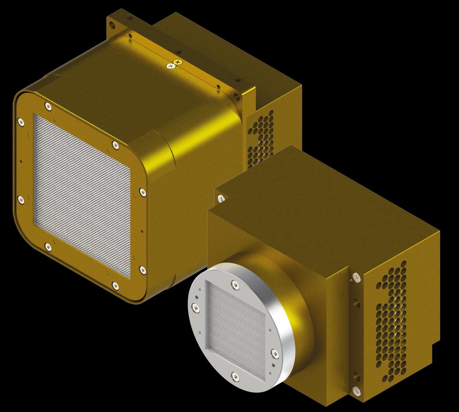
Fibre-optic coupled 4096 × 4096 X-ray detection with air or water cooling and two standard active input sizes.

Fibre-optic coupled 4096 × 4096 X-ray detection with a compact, brass-shielded housing optimised for short exposures.

Direct fibre-optic coupling protects the sensor while preserving detail in compact, high-resolution X-ray imaging.

A cooled 4-megapixel detector platform with three optical input sizes for laboratory and beamline X-ray imaging.

A large-area X-ray detector built around four fibre-coupled 16MP sCMOS sensors.

A back-reflection detector with a central beam path for real-time Laue diffraction and crystal orientation.