Back to Home
Precisometer · Official representative

Radiography systems

2D Micro Radiography — High Resolution

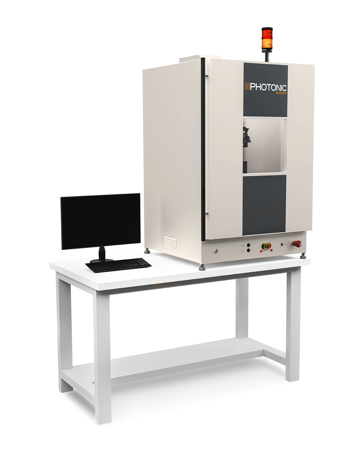
An integrated X-ray cabinet for microfocus imaging and fine detail with motorised scanning and image stitching.

2D Micro Radiography — High Resolution

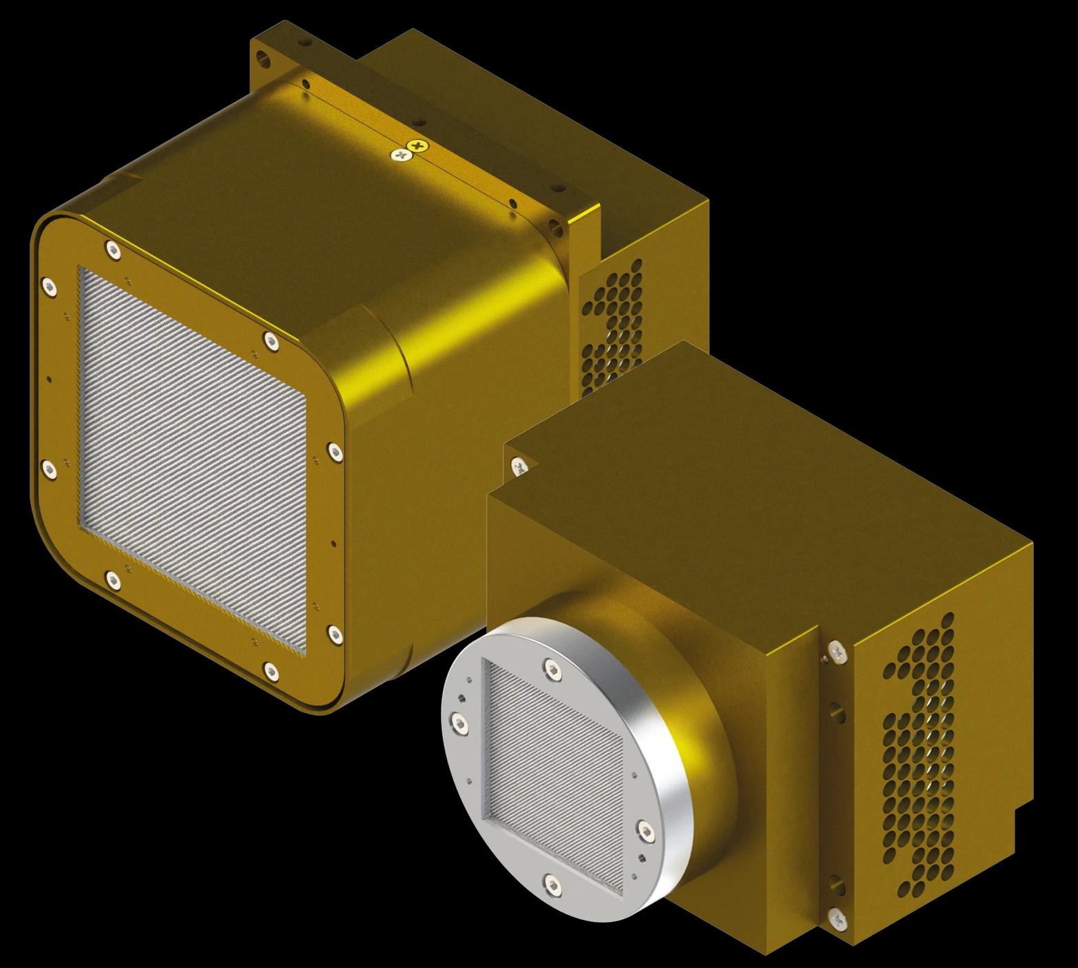
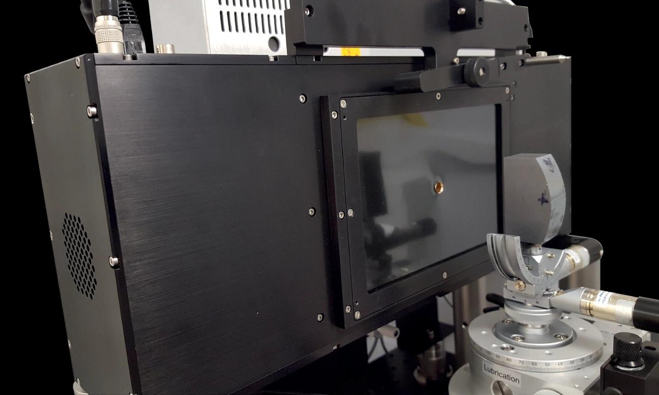
Representative manufacturer photograph; housing and connectors depend on the selected configuration. Select the photograph to enlarge.

Source
Microfocus
Object resolution
Down to 3 µm with suitable geometry
Acquisition
Motorised scanning and image stitching
Integration
sCMOS detector, motorised sample mount, alignment camera and cabinet

From photon to signal

Response for your configuration.

Full specifications

No system-level QE or DQE curve is published for this cabinet. Detector efficiency depends on the configured camera, scintillator, X-ray spectrum and sample geometry; source and detector specifications are listed below.

The details behind the selection

Full technical specifications.

Compare sensor configurations, coating options, operating limits and integration details directly below. Check the operating mode and temperature alongside each performance value.

01

Source, resolution & cabinet comparison

7 parameters
Source, resolution & cabinet comparison
ParameterHigh ResolutionRapid Imaging
X-ray source60 kVp microfocus50 kVp minifocus; 50 W
Resolution at target3–5 µm30 µm
Magnification5–10×1:1
Typical exposure in model feature listApproximately 2 s; sample dependent>1 s; sample dependent
Cabinet (W × D × H)694 × 782 × 1200 mm694 × 782 × 800 mm
CabinetFully self-shielded benchtop cabinetFully self-shielded benchtop cabinet
Rapid-imaging inputDetector selected for the applicationUp to 95 mm diagonal

The introductory text describes rapid exposures below 1 s, while the model feature list gives >1 s typical. Both are retained with their context; exposure depends on sample, source setting and detector. Resolution also depends on magnification and scintillator.

02

Sensor electronics

5 parameters
Sensor electronics
Key SpecificationsHigh-Resolution SystemRapid Imaging System
Resolution at the target3 µm at 2.5W with 5x magnification 5 µm at 8W with 5x magnification*~30 µm
Magnification range5x to 10x~1:1
Area of illumination target7.34 mm2 at 5x magnification 3.67 mm2 at 10x magnification~36.7 mm2 or ~65.5 mm2
Typical exposure time (sample dependent)~2 sec>1 sec
03

Detector head, mechanics & acquisition

15 parameters
Detector head, mechanics & acquisition
Detector Specifications*High-Resolution SystemRapid Imaging System
Number of Pixels4096 x 40964096 x 4096
FOV36.7 x 36.7 mm36.7 x 36.7 mm or 65.5 x 65.5 mm
Effective Pixel Size9 µm x 9 µm9 µm x 9 µm or 14.1 µm x 14.1 µm
Dynamic Range12000:1 with all corrections
Frame Rate4.5 fps at full sensor resolution; 18 fps at half sensor resolution
Full Well Capacity65000 to 70000 typical in mixed gain mode
Read Out Noise<5 electrons RMS at full sensor resolution and mixed gain mode <10 electrons RMS at half sensor resolution
Dark Current<1 electron/pixel/second air cooled with all corrections
Digitisation16-bit
Peak QE58% at scintillator emission wavelength (without microlens)58% at scintillator emission wavelength
Exposure50 microseconds up to 60 minutes
Spatial Resolution30 lp/mm30 lp/mm or 20 lp/mm
Detector InterfaceGigabit Ethernet
Energy Range5 keV - 55 keV with Gadox:Tb
04

Image processing & software

7 parameters
Image processing & software
X-ray SourceHigh-Resolution SystemRapid Imaging System
Power8W50W
Spot Size5 µm at 8W50 µm at 50W
Energy Range20 - 60 kVp
X-ray Beam Angle40º round beam
Target MaterialTungsten
Cooling MethodAir-cooled

Features & Benefits

  • Complete System - Fully integrated, with table, for plug-n-play operation - no additional room preparation or services required Fixed Focus Visual Camera - Aids in aligning the sample to the X-ray beam and selecting desired imaging area Motorised Sample Mount - Automated sample scanning and positioning sCMOS X-Ray Camera - Active area up to 65.5 x 65.5 mm, high sensitivity, short exposures and low noise imaging High Resolution - X-ray imaging resolution down to 3 µm FWHM with 5x magnification Air Cooled Source - Low maintenance, low power requirements - run off a standard plug Targeted magnification 1x to 10x variable magnification option for versatile systems which need flexible resolution X-ray Safety Warning lights, x-ray beacons, and interlocks conforming to Health & Safety Standards On-site/Remote Install and Training Sample 2D Radiography Images

Build your enquiry

Start with your application.

Capture the system requirements and integration details you want to discuss with Precisometer.

2D Micro Radiography — High Resolution configuration and enquiry

Tell us about your experiment so we can specify the system with you.

Your starting configuration

2D Micro Radiography — High Resolution

Source
Microfocus
Object resolution
Down to 3 µm with suitable geometry
Acquisition
Motorised scanning and image stitching
Integration
sCMOS detector, motorised sample mount, alignment camera and cabinet

Selected options are requirements for discussion. Compatibility and final performance are confirmed for your configuration.

Review enquiry

Discuss selection, integration and a quotation with Precisometer.

Technical overview

Designed around the measurement.

An integrated X-ray cabinet for microfocus imaging and fine detail with motorised scanning and image stitching.

Non-destructive testingMaterials scienceElectronicsArchaeology
2D Micro Radiography — High Resolution technical overview
SourceMicrofocus
Object resolutionDown to 3 µm with suitable geometry
AcquisitionMotorised scanning and image stitching
IntegrationsCMOS detector, motorised sample mount, alignment camera and cabinet

Explore the wider range

Other routes to your measurement.

All Photonic Science products
Product selection

What do you need to measure?

Start with your signal, or search directly for a camera, sensor option or application.

6 product ranges · 16 camera configurations

X-ray sCMOS FDS
Photonic Science / X-ray detectors

X-ray sCMOS FDS

Direct fibre-optic coupling protects the sensor while preserving detail in compact, high-resolution X-ray imaging.

Standard configurations
FDS 1.77MP_17_S, 2.86MP_11_S and 7.01MP_17_S
Acquisition
10–37 fps, model dependent
3 configurations · specifications
X-ray sCMOS 4MP
Photonic Science / X-ray detectors

X-ray sCMOS 4MP

A cooled 4-megapixel detector platform with three optical input sizes for laboratory and beamline X-ray imaging.

Resolution
2048 × 2048 pixels
Input sizes
22.5, 48 or 67.2 mm square
3 configurations · specifications
X-ray sCMOS 64MP
Photonic Science / X-ray detectors

X-ray sCMOS 64MP

A large-area X-ray detector built around four fibre-coupled 16MP sCMOS sensors.

Architecture
Four 16MP sCMOS sensors with fibre-optic coupling
Acquisition
Over 5 fps full resolution; subarea / line modes available
1 configurations · specifications