Automated brightfield slide scanner
Digitise microscope slides at 20× or 40× optical magnification, with capacity for up to 320 slides.
Technical summary with specifications and configuration choices.
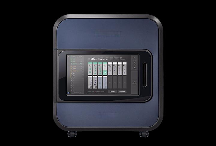
Features
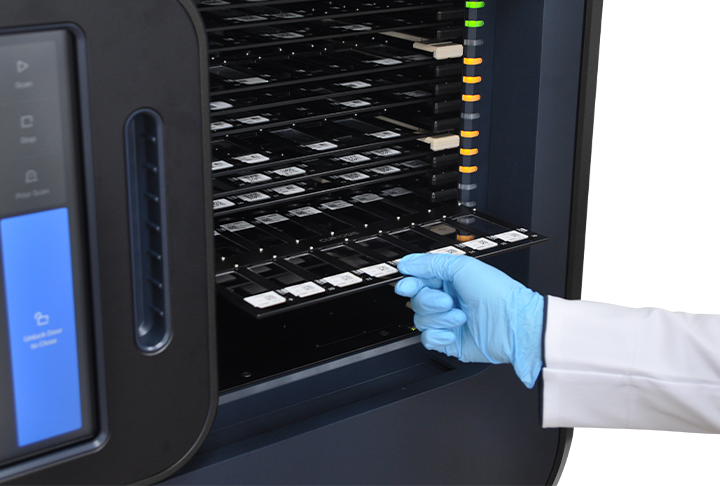
- Automated loading of up to 320 slides, with continuous tray loading during scanning.
- 20× and 40× optical scanning options for different sampling requirements.
- Focus-map acquisition, image stitching and priority tray scanning.
- Standard and optional mega-slide trays accommodate different specimens.
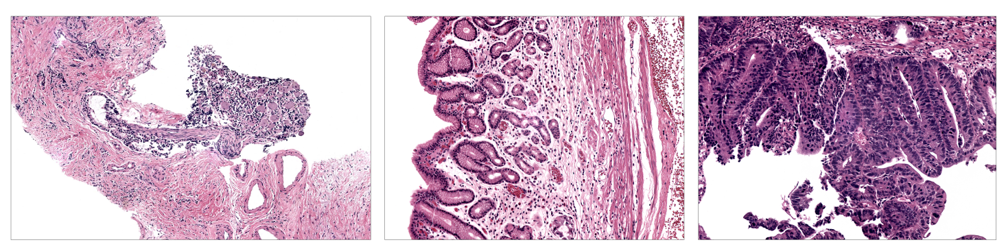
Images and workflow examples
Select an image to view it at full size.
Technical specifications
| Slide capacity | 320 slides, 20 trays × 16 slides |
|---|---|
| Optical magnification | 20× or 40× |
| Sampling at 20× | 0.225 µm/pixel |
| Sampling at 40× | 0.112 µm/pixel |
| Scan time at 20× | 14 s for 15 × 15 mm tissue |
| Scan time at 40× | 23 s for 15 × 15 mm tissue |
| Field of view | 20× 0.72 × 0.50 mm; 40× 0.36 × 0.25 mm |
| Focus | Focus map |
| Standard scan area | 24 × 54 mm |
| Optional mega scan area | 44 × 54 mm |
| Standard slide size | 24–25 × 74–76 mm |
| Optional slide size | 50–52 × 74–76 mm |
| Label area | 25 × 15–20 mm |
| Scan mode | Brightfield |
| Touchscreen | 52.1 × 29.3 cm |
| Dimensions | 82.5 × 72 × 91.5 cm; 116 × 72 × 91.5 cm with door open |
| Weight | 175 kg |
| Power input | 220–230 V AC, 50/60 Hz |
| Operating temperature | 10–35 °C |
Applications
- Digitise stained microscope slides for research review and image analysis.
- Batch acquisition of standard and selected large-format tissue slides.
Configurations and accessories
- Specify 20× or 40× scanning, standard or mega trays, and the required slide formats.
- Include workstation, image storage, network integration and available installation space in the enquiry.
Selection notes
- Specifications follow the current revision-6 brochure. Older webpage figures differ. Confirm magnification, sampling and power input for the selected configuration; scan times refer to the stated tissue area.
Discuss your experiment
Tell us what you need to measure. Next, add your contact details and review the enquiry before sending.